
 The Immune System:
The immune system represents a family of biological molecules found inside every living organism, dedicated to provide protection to the host against any foreign entity, be it pathogenic microbes, particulate matters from unknown sources or even cells (tissues, organs) from a donor (during replacements, transplants and grafts). The basic of this defence mechanism lies in the recognition of the entity by the immune system as ‘known’ or ‘self’ (native to the host) and ‘unknown’ or ‘non-self’ (foreign to the host). The immune system identifies the ‘self’ and non-self’ molecules on the basis of markers present on their surfaces. The ‘non-self’ markers help the immune system in identifying foreign bodies, which are biologically termed as antigens. It is these antigenic markers (referred to as epitopes) trigger an immune response in a healthy host, leading to a cascade of events, resulting ultimately in the destruction of the same.
A human body has its own set of ‘self’ markers in the form of a group of proteins known as the Major Histocompatibility Complex (MHC) which is unique to each individual. The MHC is responsible for presenting total or a part of the foreign antigen to the immune system, following which the immune cells trigger a reaction favouring the host and clearing the foreign entity. There are two main classes of MHC molecules, out of which MHC class I molecules are found on most nucleated cells except red blood cells (RBC) and MHC class II molecules are found on the surface of a specific class of immune cells collectively called Antigen Presenting Cells (APCs). The basic mechanism involves taking up the pathogen by the immune cells (MHCII) or infected cells (MHCI) and processing of the same, followed by presentation of part of the antigenic molecule in the form of markers (epitopes) in association with the MHC molecules, which presents itself along with the epitope on the cell surface, only to be recognized by special immunizing structures (antibodies) for eventual destruction.
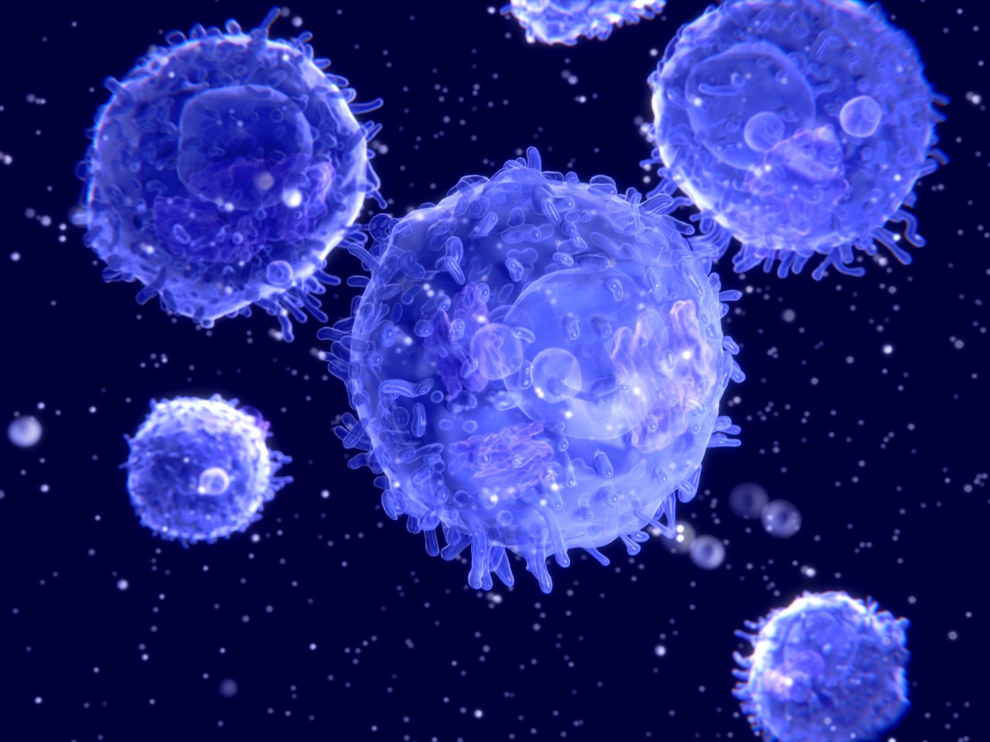
The major immune cells include lymphocytes or white blood cells (WBC) which include Natural Killer Cells, Macrophages, Dendritic cells, T lymphocytes and B lymphocytes, which originate from the lymphoid organs in the body. These organs include the thymus, the spleen and bone marrow, along with a system of fine networks connecting these organs to each other, called the lymphatic system. Lymph runs through these vessels, providing nourishment to these tissues. These vessels (lymph vessels) have their openings called lymph nodes at various organs of the body (including the armpits, neck, abdomen, chest and groin among others) where there are special compartments dedicated for immune cells to attack and eliminate antigens. The incoming lymph vessels act as the entry for the antigens and newly synthesized immune cells, and the outgoing lymph vessels aid in their exit. Once released into the bloodstream, the now matured immune cells are spread throughout the body where they interact with pathogens in a more structured manner, with specific cells having specific roles towards providing immunity to the host.
The bone marrow is responsible for synthesis of hematopoietic stem cells which are the most primitive progenitors of all kinds of cells operating in the human body. They give rise to all kinds of cells including blood cells and immune cells. The two main categories of cells formed from stem cells are myeloid and lymphoid progenitor cells, out of which the myeloid progenitor cells give rise to immune cells which are mostly generic in nature and form the first level of defence mechanism needed for the body on attack by pathogens, and the lymphoid progenitor cells are involved in formation of specific mechanisms to get rid of the pathogens already presented by the APCs.
 The common immune cells arising from the myeloid progenitor cells include mast cells, natural killer cells, eosinophils, neutrophils, basophils, macrophages and dendritic cells. Their main role lies in identifying a foreign pathogen with the help of pattern recognition receptors (PRRs, a group of receptors which help in identifying pathogen based molecular patterns associated with different microbes, stress caused to the cell in any form or damage caused to cells. A few such examples include Toll-like receptors, Receptor Kinases and C-type lectin receptors among others), engulfing the same, sending out warning signals which result in the release of chemical compounds and messengers like histamine, serotonin, bradykinins and prostaglandins and chemokines that dilate blood vessels causing inflammation and pursue compartmental killing of pathogens, a process known as phagocytosis. Characteristically neutrophils are the first cells to arrive at the sight of a foreign pathogen and engulf it and release free oxygen radicals, peroxide and hypochlorite, followed by macrophages which are the most efficient in terms of phagocytosis (their receptors bind to those present on the surface of the antigen, mostly bacteria in this case, and release chemicals to kill the entrapped pathogen), followed by recruitment of granular cells like basophils, eosinophils and mast cells, which are related to production of histamines, heparins and activating chemokines and cytokines (the latter two being chemical messengers leading to specific immune responses after triggering a cascade of events collectively called the complement system). The dendritic cells being located on the mucosal lining of most organs and tissues lining the skin, are mainly associated with antigen presentation to the other immune cells after interaction with the pathogen followed by binding to specific MHC molecules. These chains of events collectively constitute the innate immune system. They further lead to specifically tailored immune responses in most cases, a branch of immunity known as adaptive immune system.
The lymphoid progenitor cells are responsible for giving rise to the bearers of acquired immunity in the human body – an event which creates an ‘immunological memory’(ability to distinguish similar ‘self’ and ‘non-self’ markers in the future) within the host which equips the same with the ability to combat similar infections in future. The T-lymphocytes and B-Lymphocytes are the main cells associated with adaptive immunity, where the T-cells mature in the thymus and the B-cells mature within the bone marrow itself. T cells are mostly associated with providing cell-mediated immunity whereas the B cells are concerned with the humoral (pertaining to body fluids) branch of adaptive immune responses. This mode of immunity (acquired mostly through vaccinations as a measure to protect against severe infections) is called so because of a series of beneficial genetic mutations that take place within the genetic make-up of most antigen receptors, unique to each of the trillion lymphocytes concerned with this, which when divide and give rise to progenies, have the same beneficial gene combination. Since these mutations are irreversible, the gene code of the parental DNA gets conserved in the offspring. The result of this is the formation of memory T cells and B cells which are the most effective in providing long term immunity against targeted infections.
T cells can affect the immune system in two ways: firstly, there are cytotoxic variants of these lymphocytes (cytotoxic T lymphocytes or CTLs) which attack virus or bacteria infected cells directly, and helper T lymphocytes which help in managing the overall immune system, helping in accentuating and enhancing the performance of APCs and B cells. The newly synthesized CTLs with their T cell receptors (TCRs) look for a (antigenic) peptide bound MHCI complex. On binding of the TCRs to the MHCI and peptide complex, the CTLs become more potent and active, releasing cytotoxins (like perforin and granulysin) causing lysis of the infected cells, and then spread throughout the body via body fluids in search of similar MHCI and peptide combinations. On the other hand, the helper T cells (Th) do not possess any cytotoxic or phagolytic ability, but help in the secretion of chemical messengers (chemokines and cytokines) which in turn activate a cascade of events collectively called the complement pathway, which eventually helps in enhancing the potential of the APCs and B cells. The TCRs on these Th cells bind to MHCII molecules bound to antigenic peptides.
 B lymphocytes are involved in humoral immunity and work mainly via the synthesis of antibodies. They are distinguished from the other lymphocytes due to the presence of the B cell receptor (BCR) protein on their surface, which is involved in recognition of appropriate antigens as presented by the macrophages and helper T cells. Another basic difference between antigen recognition by T cells and B cells is that B cells can recognize its antigen in its native form in the body fluids with the help of the BCR whereas cytotoxic T cells need MHCI molecules bound to the antigen for them to act upon. B cells however perform better when helper T cells join them in antigen processing. The B cells are in a state of constant synthesis in the bone marrow (in millions) and are naive when newly synthesized. Once exposed to antigens in the body fluids, they transform into plasma B cells, and secrete antibodies specific to the antigens they combat, at a rate of up to 10 million copies an hour (which is why they are often termed as ‘antibody factories’). These cells are short lived and die once the target antigen has been dealt with. Some of these cells transform into memory B cells which are long lived and recognise the specific antigenic epitope it is supposed to bind to. The number of memory B cells increases with every cycle.
(Antibodies secreted by the B cells belong to the Immunoglobulin super family of proteins and are basically a ‘Y’ shaped molecular structure with a constant region corresponding to the stalk of the ‘Y’ shaped structure, and the two arms corresponding to the variable region with a unique genetic makeup, as sorted out during maturation and differentiation of plasma B cells, destined to attack a particular antigen. Out of the immunoglobulins (Ig) present in our body, IgG is present in majority in blood, being the only immunoglobulin to be transferred from the maternal circulation to the fetus via the placenta, and is effective in killing most bacteria and microbial pathogens, IgD is involved in B cell activation and is found on the surface of the B cells, IgE is responsible for binding to allergens and helps the basophils and mast cells in releasing histamines and other neuro-inflammatory compounds, IgA acts as a immune barrier at the common entry points of the body, embedded in body fluids like tears, saliva, secretions of the respiratory and gastrointestinal tracts, and IgM being a constant member of the bloodstream, effectively eliminates microbial pathogens before enough IgG is secreted.)
B cells (antibodies bound to antigens), along with a few of the APCs secrete chemical messengers and hormones like chemokines and cytokines in order to activate the complement system. This system comprises of small proteins (around 25 variants) secreted in the liver which act as precursors in antibody mediated killing of pathogens. These proteins include serum proteins, serous proteins (from fluids found inside body cavities) and cell membrane receptor proteins. They have different biochemical variants as far as disease specific pathways are concerned.
It is through the combined action of all these components of the immune system that the healthy host combats infections and pathogens.
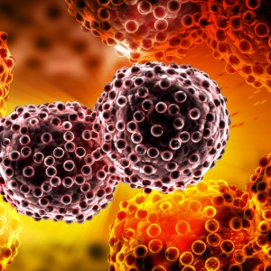 Immune System and Cancer:

Impairment of the immune system due to any reason (radiation therapy, infection, transplants, trauma or congenital deficiencies) gives rise to a number of disorders, which include autoimmune diseases (when the immune system is unable to distinguish between ‘self’ and ‘non-self’ proteins and hence targets cytotoxic T cells against it’s own host), allergy (where exposure to allergens increases production of IgE immunoglobulins leading to increased inflammation reaction), acquired immune deficiency disorder (AIDS: when one or more components of the immune system, mostly T helper cells, go missing or get depleted, mostly due to infection with harmful viruses), immune complex disease ( due to accumulation of antigen-antibody complexes which remain trapped within tissues instead of getting eliminated, and hence cause damage to the organs nearby), common variable immunodeficiency (CVID: which represents a condition with multiple autoimmune disorders together, like inflammatory bowel disease, autoimmune thrombocytopenia and autoimmune thyroid disease) and cancer, being another prominent consequence.
Recent research over the past two decades has shed considerable light upon the fact that apart from protecting the host from different kinds of infectious pathogens, the immune system has a major role in framing and modelling tumour immunogenicity. The entire process is known as cancer immunoediting which consists of three phases namely elimination, equilibrium and escape.
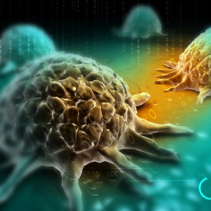
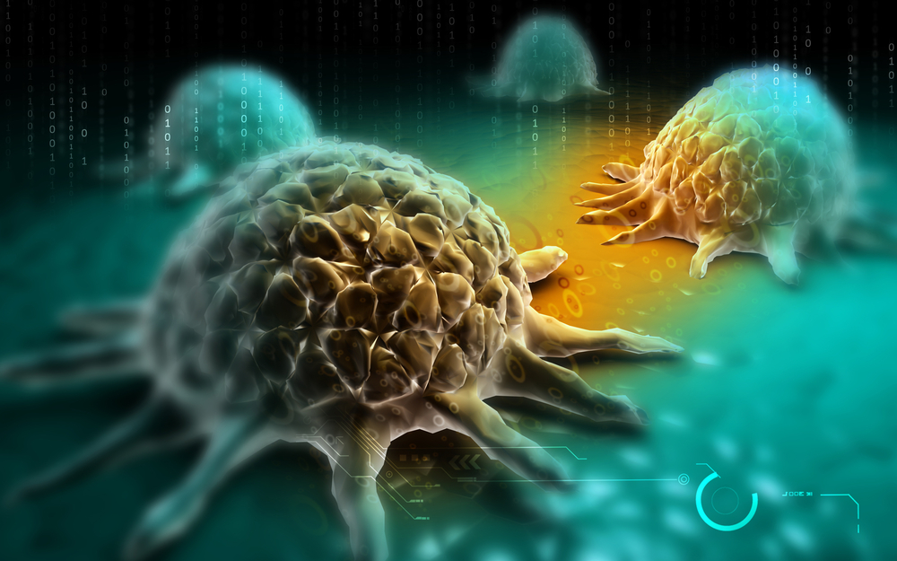
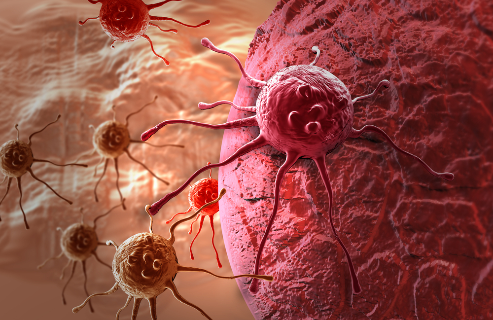
- Elimination: This phase is also known as cancer immunosurveillance and refers to a collective group of innate and adaptive immune responses exerted by the immune cells to the mass of cells which are most probable to give rise to a tumour. When normal cells turn to tumour cells, some of the antigens on their surface change and they shed some of these proteins in the bloodstream, which are often tumour antigens. The cells of the innate immune system are recruited first in this case after inflammatory signals are sent out. Natural Killer (NK) cells, NK T lymphocytes, macrophages and dendritic cells are normally the main ones infiltrating these tumour cells. They engulf these affected cells and secrete cytokines, Interferon gamma (IFN-gamma) being the most common of them. This induces death of some of the tumour cells and secretion of several other chemokines like CXCL 9, 10 and 11, which help in preventing growth of the tumour by blocking blood vessels, which eventually stop the flow of nutrients necessary for the tumour cells to grow. This causes a gradual death of these cells which are later engulfed by dendritic cells which migrate towards the outgoing lymph nodes. This process continues as more immune cells are recruited by the circulating chemokines and more and more tumour cells are eliminated from the healthy host. The dendritic cells trigger the production of more cytotoxic T lymphocytes which eventually destroy these tumour cells. Apart from this, the macrophages and NK cells act mutually in activating each other further to induce apoptosis of the tumour cells (apoptosis, referring to the programmed cell death according to a normal cell cycle) by secreting more IFN-gamma and IL2 (Interleukin 2). This enhances he secretion of perforins, free radicals and TNF-related apoptosis inducing ligands (TRAILs) which aid in cytotoxicity. The role of NK cells in tumour immunosurveillance has been highlighted in a number of studies and shows that several receptors on the surface of the NK cells play a pivotal role in recognizing tumour antigens and tumour specific proteins released in the bloodstream. Some of these receptors include NKG2D, NKp30, NKp44 and NKp46, all of which induce a higher potential of cytotoxicity within these cells. Mouse studies with NK cells have also shown a higher incidence of tumour cells and greater chances of malignancy in NK deficient mice. These prove the importance of NK cytotoxic cells in tumour immunosurveillance, apart from the other components of the immune system.
- Equilibrium: Despite attempts made by the immune system to monitor the growth and development of tumour cells, there are a lot of cells which escape this screening mechanism and proceed to the equilibrium stage. They are supposed to have a non-immunogenic phenotype and their growth in this phase lasts for around a few years – the longest phase of the three. Several theories suggest that natural selection (a Darwinian concept) acts upon these budding tumour cells and eventually those with the maximum potential to combat the immune system, survive (mostly because of induced mutations developed in course of time, owing to their abnormal growth pattern, that aid in their survival among the cytotoxic cells of the host.)
- Escape: This is the last phase of immunoediting where the mutated tumour cells continue to grow and eventually invade the host’s immune system through various mechanisms and lead to malignancies and cancer.
The tumour cells can escape immunesurveillance mechanisms in a number of ways. Some of them might mimic MHCI molecules and escape attack by the APCs and invade the immune system of the host. Others might express mutated forms or soluble forms of proteins and markers on their surface which makes it difficult for NK cells and their receptors to identify the appropriate tumour cells. One such example is the expression of the receptor NKG2D on NK cytotoxic cells which identifies NKG2DL ligand expressed by the tumour cells (mostly caused as a result of stress, genetic toxicity, DNA damage, viral infection and heat shock among others, and is a major component of precancerous lesions and many tumours in human hosts). Research has shown that the amount of NKG2DL markers can be directly be correlated to determine the capacity of the immune system to monitor tumour growth and stop the same. Memory T cels have also been seen to have formed after interaction of cytotoxic lymphocytes and NKG2DL interactions. However, some tumour cells send out inhibitory signals which counters the activation of NK cytotoxic cells despite the presence of NKG2DL on their surface, hence the NK cells are unable to perform at the potential they are expected to. Another explanation comes in the form of soluble proteins released by the tumour cells which mimic antibodies and hence help in escaping tumour immunosurveillance and allow advancement of the tumour. Another theory suggests that tumour cells produce immunosuppressive cytokines like Tumour Growth factor Beta (TGF-beta) which suppresses the activity of the antigen binding receptors on the surface of NK cells, hampering their activity. Apart from these a number of different mechanisms have been hypothesised by authors to show the different means adapted by tumour cells to combat the immune system and lead to malignancies.
 Cancer Immunotherapy:

Once a person is diagnosed with cancer, which leads to formation of an uncontrolled mass of ill developed cells, it becomes necessary to kill these abnormal cells in order to restore normal conditions. Unfortunately one has to undergo a series of painful procedures to achieve the target of eradicating the cancerous cells, that too, not being 100% efficient. This is because the common methods used to treat cancer include irradiation (burning the malignant cells), chemotherapy (poisoning the cells) and surgery (removing the cells from the affected organs, but that is possible only during the early stages of the disease). These methods are however not very dependable with no sure shot results. The chief reasons behind this being health issues of patients concerned. A majority of the available treatment options, whether applied as monotherapy or in combination, have a tendency to be toxic for healthy cells as well, killing them in as many numbers, as the cancer cells. This often takes a toll on patients who fall within the older age group, who have comorbidities and health issues as well. Immunesuppression is another unwanted effect of chemotherapy and radiation sessions.
Till date there has not been a ‘fool proof’ strategy to combat this deadly condition, but scientists have been working on ways which might utilize the body’s immune system to combat cancer, with parallel therapy as and when needed. A simple logic for this approach lies in the fact that the immune system is the only natural and the least toxic tool for fighting any kind of disease within the human body. Normally an external stimulus is applied to the immune system of the patient as it is already weakened and needs a ‘trigger’ to work at its full potential or even more successfully, as the situation demands. Another method is to inject some of the components of the immune system cultured artificially in the laboratory with a higher potency with tumour derived proteins, so that these when administered within the human system, can recognise the cancer cells as foreign bodies and destroy them. Hence, in a nutshell, immunotherapy can be referred to as a biological therapy to effectively treat cancer in a non-toxic way as compared to conventional treatment options.
There are three main classes of immunotherapy, which involve cell based, antibody based and cytokine based methods. These involving monoclonal antibodies and cytokines are collectively termed as passive immunotherapy. Administration of immune cells (modified to suit the severity of the disease) via vaccinations is referred to as active immunotherapy.
- Active Immunotherapy: This method involves two main techniques, a) Cell based methods, where a person’s own cells are used to treat the patient’s cancer, and b) vector based methods, where an external vector is genetically engineered in the laboratory to express tumour specific antigens and then infused with immune cells for injection.
- Cell based method: This method acts as a ‘booster’ to the patient’s immune system by injecting potent immune cells directly into the bloodstream. It not only triggers the immune system as a whole but also provides the ability to combat cancer cells. The most commonly applied therapy in this category involves dendritic cells. They are one of the most potent APCs and form a link between both innate and adaptive immunity, which makes them strong contenders for this kind of an approach. As we know, dendritic cells have the ability to induce a massive T cell mediated immune response when presented with an antigen. This is the main idea behind development of dendritic cell based vaccines. Dendritic cells can be extracted from a patient’s own body, and converted to a recombinant protein by infusing along it, with some of the tumour peptides which correspond to those present on the surface of cancer cells, along with adjuvants which enhance the formation of more immune cells once inside the body and boost the immune system (one such adjuvant is the Granulocyte Macrophage Colony Stimulating Factor, or GM-CSF, which is a cytokine that enhances the production of more white blood cells and ensures their differentiation and maturation into eosinophils, neutrophils, basophils, monocytes and mast cells). This also helps in producing avery reliable anti-tumour response within the immune system. This modification can also be done within the patient’s body, by genetically inducing the tumour cells to produce and secrete GM-CSF. Sometimes Interleukin 2 can also be administered to provide better results as it regulates the activities of the white blood cells and ensures it distinguishes ‘self’ and ‘non-self’ proteins effectively. Once this modification has been achieved, these recombinant dendritic cells trigger a massive inflow of cytotoxic T-cells which have the potency to perform immunoediting, and in turn, eliminate the cancer cells. More modern techniques use antibodies specific to the receptors on the surface of the dendritic cells. Tumour antigens (proteins) can then be added to these antibody coated receptors in order to provide long term protection against the tumour cells of the patient. Some of the common receptors used in this case include Toll Like Receptor (TLR) 3, 7 and 8 and CD40.
- Vector based methods: This method is a modification of the in-vitro and in-vivo methods used in order to make a recombinant APC (dendritic cell). Here, a viral vector is used to transfer the tumour antigens and combine them with the dendritic cells. They are cultured artificially under suitable growth conditions in the incubators and are made to express GM-CSF or other adjuvant within their genetic material. This is then combined with the dendritic cells and the entire recombinant (protein) is then ready to be injected into the patient’s body.
One therapeutic vaccine which was approved by the UD FDA in 2010 is Provenge (Sipuleucel-T) which is used to treat minimally symptomatic, hormone refractory metastatic prostate cancer and has been shown to increase the survival by a median of 4.1 months (according to phase III clinical trial reports). It involves injection of APCs from the patient’s body and infuse it with a fusion protein PA2024, consisting of, PAP protein (Prostatic Acid Phosphatase) which is present on most prostate cancer cells, and GM-CSF, into a recombinant protein (Activated Blood Product, APC8015) which is then injected back into the patient. It is repeated three times each with a gap of two weeks in between, and the entire procedure is prostate specific since it has the PAP protein. One disadvantage of this technique lies in its cost, which is as high as USD 93000. Affordability becomes a big question in this case and when one compares it to the results, one might think twice before opting for it.
- Passive Immunotherapy: This method mainly employs the use of monoclonal (laboratory made, similar in nature) antibodies and cytokines and chemokines to provide protection against specific or single targets within the patient’s body. They can be directed against a particular antigen or a particular protein on the surface of the cancer cells or a cancer specific enzyme or protein. On one hand, antibodies are one of the most potent candidates because of their role in adaptive immune responses and being specific for diseases and also for being the most effective in destroying the harmful effects of antigens. On the other hand, cytokines and chemokines are known for their role in modulating immune responses, because of which they can be utilized as useful candidates to treat cancer cells.
- Antibody based therapy: Monoclonal antibodies are created in a laboratory and often involve mouse models, the antibodies thus formed being known as murine antibodies. However, when mouse cells are used there is always a risk of immune rejection and harmful side effects when injected within the cancer bearing host. Chimeric antibodies, which consist of part of mouse and part of human antibodies are created to counter the risk of adverse immune reactions caused as a result of their murine counterparts. Humanized antibodies are mostly derived from human antibodies with only a few parts of it being replaced by their murine counterparts. Human antibodies are completely derived from humans with similar amino acid sequences.
 The method of production of monoclonal antibodies involves the introduction of antibody producing plasma cells and infusing and immunizing them with a cancer specific antigen within a suitable host (be it mouse or human) and later combine them with a long-lived cancerous immune cell called myeloma cell. This combination is screened for the desired antibody production using selective media and on obtaining the requisite antibody, they are tested for their capacity to bind to a desired antigen using molecular biology assays (like ELISA and immune blotting technology). The most suitable combination (now called a hybridoma) is then cloned so that they form similar antibodies. Hence these are named ‘monoclonal’ antibodies. These antibodies are normally specific to a single type of cancer cell as one single type of antigenic protein is used to make them.
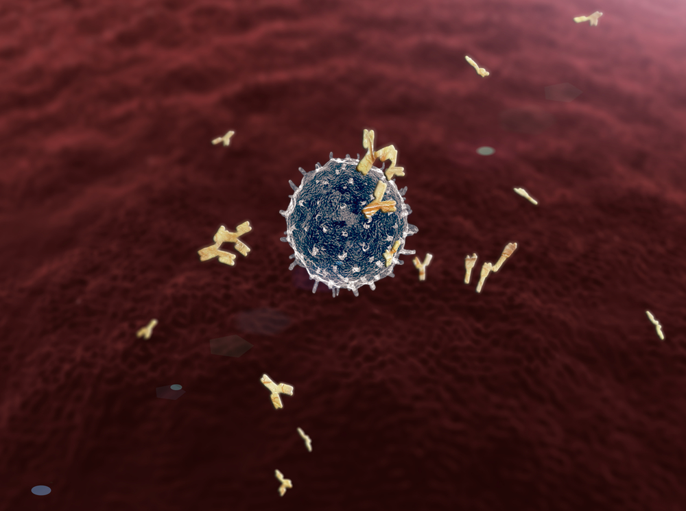
The monoclonal antibodies have different ways in which they attack cancer cells. One such pathway is called the Antibody Dependent Cell-Mediated Cytotoxicity (ADCC). This needs the antibody to be bound to the surface of the target cells (here, the cancer cells). The constant region of the antibodies bind to the receptors for these constant regions (denoted as Fc regions and Fc receptors respectively) on the surface of immune cells, especially the NK cells, upon which they release their enzymes needed for programmed cell death of the tumour cells (the tumour cells bind to the variable regions of the antibodies, Fab, meant specifically for specific antigen binding).
Complement system mediated cell killing is another method by which destruction of tumour cells is achieved. Chimeric, humanized or human antibodies can be used in this case, if they contain the IgG1 based Fc region (of the Immunoglobulin superfamily of proteins present in humans). The complement system gets activated when the antibody binds to the target tumour cell, and components of the complement pathway (be it classical or alternate) engulf the target cell to form a Membrane Attack Complex (MAC). These components then perforate the membrane of the target cells and cause lysis. This leads to the eventual destruction of the tumour cells. On the other hand, it can also accentuate the ADCC pathway and help in antibody mediated killing of tumour cells.
Cell signalling is another key method of antibody mediated killing of tumour cells. When the monoclonal antibodies bind to useful receptors on the surface of blood cells or the cancer cells themselves, which are meant for vital cell signalling pathways needed for survival of these cancer cells, they block these binding sites which otherwise would have been bound to other external proteins, peptides or small molecular compounds called ligands. Because these ligands bound to receptors on the tumour cell surface are essential for sustaining the cancer cell population, antibodies bound to these receptors act antagonistically and help in stopping their (cancer) cell cycle, inducing apoptosis. This category of antibodies is known as antagonists. The apoptotic pathway is one of the majorcell signalling pathways utilized by monoclonal antibodies in order to kill cancer cells.
Some of the antibodies approved by the US FDA include:
- Avastin, chemically known as Bevacizumab, a humanized IgG1 monoclonal antibody which binds to vascular endothelial growth factor-A (VEGF-A), and uses apoptotic cell signalling pathway to bock the growth of tumour cells. This has been recommended for colon cancer, kidney cancer, breast cancer, lung cancer and glioblastoma, among a range of various countries.
- Alemtuzumab (Campeth-1H), an anti-CD52 humanized IgG1 monoclonal antibody indicated for the treatment of fludarabine-refractory chronic lymphocytic leukemia(CLL), cutaneous T-cell lymphoma, peripheral T-cell lymphoma and T-cell prolymphocytic leukemia. This works on the basis of the ADCC pathway of tumour cell killing.
- Erbitux, chemically known as Cetuximab, is a chimeric IgG1 monoclonal antibody that targets the extracellular domain (part of the receptor outside the cell) of the epidermal growth factor receptor (EGFR). It is used in the treatment of colorectal cancer and head and neck cancer. This is another antibody which works on the principle of binding to receptors to block the binding of ligands and promote cell death.
- Rituxan, chemically known as Rituximab, a chimeric monoclonal antibody against the protein CD20, which is primarily found on the surface of immune system B cells. It destroys B cells and is therefore used to treat diseases which are characterized by excessive numbers of B cells, overactive B cells, or dysfunctional B cells. This includes many lymphomas, leukemias, transplant rejection, and autoimmune disorders. This also works on the basis of the ADCC pathway of cell killing.
- Trastuzumab is a monoclonal IgG1 humanized antibody specific for the epidermal growth factor receptor 2 protein (HER2). It received FDA-approval in 1998, and is clinically used for the treatment of breast cancer. HER-2 is a member of the epidermal growth factor receptor (EGFR) family of transmembrane tyrosine kinases. Once activated on cancer cells it activates cell signalling pathways that promote cell proliferation, cell growth, angiogenesis and metastasis, and inhibits programmed cell death (apoptosis). Trastuzumab inhibits binding of HER-2 to cancer cells and thereby induces apoptosis (cell signalling).
- Cytokine based therapy: Cytokines have the ability to modulate immune signals and hence scientists have taken the benefit of this property to use it against the tumour cells to induce cell death. The most common cytokines are Interferons (IFNs) and Interleukins (ILs). Interferons are normally produced by the body to act against viral infections, they have also shown promise to be used as therapeutic agents in order to treat cancers, especially, hairy-cell leukaemia, AIDS-related Kaposi’s sarcoma, follicular lymphoma, chronic myeloid leukaemia and melanoma. Among the three types of interferons, only IFN type 1 (including IFN alpha and beta) have been shown to be effective in cancer treatment.
On the other hand, interleukins have been shown to promote the health of T cells (IL-2) and is known as a T cell growth factor. This stimulates cytokine production and activation of multiple kinds of immune cells, which eventually boost the immune system. This therapy has been tested in treatment of renal cell carcinoma and malignant melanoma, till date.
Further Research and Development:
Since immunotherapy is a comparatively new topic to be touched upon, it has scopes for a lot of further research. Some other kinds of immune cells might be tested to add to the passive immunotherapy methods, which now focus upon only antibodies and cytokines. Using T cells to treat cancer cells by hybridizing them in laboratory conditions with tumour antigens and subsequently aiding in their activation and proliferation with the help of Interleukin 2, is a possibility being explored for the near future. Antibodies targeting several other receptors like the GD-2 protein and the CD-47 protein, expressed on the surface of different cancer cells, are potent targets of immunotherapy. Blocking of immune checkpoints inside the body, which are often used by tumour cells to escape immunesurveillance, can also be used as potential targets to monitor and inhibit the growth of tumour cells.
[amp-optin id=’13477′]