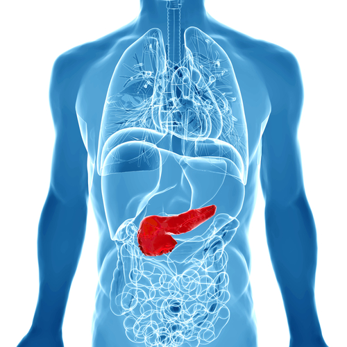
In a new study entitled “Use of 18F-2-Fluorodeoxyglucose to Label Antibody Fragments for Immuno-Positron Emission Tomography of Pancreatic Cancer” researchers developed new probes combining 18F-2-deoxyfluoroglucose (FDG) with antibody fragments, for positron emission tomography (PET) enabling a better visualization method for tumor screening. Researchers hope the newly developed probes will help clinicians achieve a more accurate and efficient detection of tumors, which could help determine patients’ clinical response to therapeutics. The study was published in the journal ACS Central Science.
Tumor cells are characterized by higher proliferation rates and, to keep this division process, they need more “fuel,” i.e. sugar, when compared to normal, healthy cells. Positron emission tomography (PET) is an imaging technique that identifies the areas within the body that are metabolically active, i.e., those areas that are consuming more sugar, for example, and that are usually associated with cancer. This is achieved by injecting a radioactive drug (tracer) that will then accumulate in these hyperactive areas. PET scans are usually performed with a labeled sugar molecule called 18F-2-deoxyfluoroglucose (FDG) to detected tumor areas. However, advancements in PET imaging has been halted due to the unstable properties of the reporter molecule, making it difficult to attach to other potentially useful molecules, besides FDG. This is particularly important, since despite tumor cells exhibit an increased FDG uptake, this molecule can actually accumulate in other non-tumorigenic but highly metabolic areas, and thus is not tumor-specific.
In this study, researchers devised a strategy to overcome these limitations generating 18F-labeled antibody fragments, i.e., linking a labeled FDG molecule to an antibody fragment. The team tested their new method and was able to detect pancreatic cancer tumors that due to their small size remained undetected using FDG molecule alone.
The team highlights that the newly developed probes can be used along with current techniques allowing a more accurate and efficient labelling of tumors based on their increased metabolic activity when compared to the healthy surrounding tissue.


