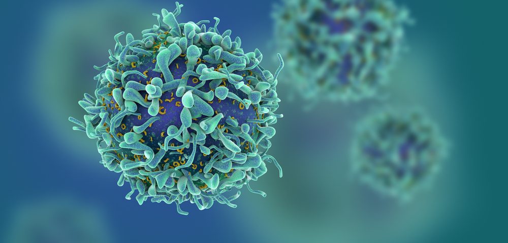
Cancer cells release into the bloodstream small vesicles armed with PD-1 ligand (PD-L1) — a protein that suppresses the activation of cancer-fighting immune cells — so as to mute anti-tumor immunity throughout the body, researchers report.
Study results also showed that vesicle-associated PD-L1 in the blood may work as a treatment response biomarker, helping to distinguish patients most likely respond to anti-PD-L1 therapies.
The study, “Exosomal PD-L1 contributes to immunosuppression and is associated with anti-PD-1 response,” was published in the journal Nature.
A well-known and powerful mechanism of immune evasion by cancer cells is the presence of PD-L1 on their cell surface, which will bind to its PD-1 receptor on T-cells to prevent the activation of T-cells — a type of white blood cell that fights cancer.
Previous studies have also shown that the presence of PD-L1 on cancer cells’ surface can increase in response to the release of interferon gamma (IFN-γ) — a protein with anti-cancer properties — by activated T-cells.
Several approved anti-cancer therapies target and block this interaction (PD-L1 with its PD-1 receptor) to promote T-cell activation and stimulate anti-canter immune responses. However, many patients with PD-L1-producing cancers do not respond to these therapies.
A collaboration between researchers — those at the University of Pennsylvania, Wuhan University, Xi’an Jiaotong University, Wistar Institute, University of Texas, and Mayo Clinic — sheds light on the immune-evasion mechanisms of cancer and possible causes behind the non-response to therapies.
The researchers found that human melanoma cells not only have PD-L1 on their surface, but they also release into the bloodstream large amounts of small vesicles — called exosomes — with PD-L1 on their surface, which systemically suppress anti-cancer immune responses. This finding was also true for breast and lung cancer cells.
In a mouse model of melanoma, injections of PD-L1-vesicles promoted tumor growth and led to a significantly lower number of T-cells able to infiltrated tumor cells and the lymph nodes. The addition of antibodies against PD-L1 strongly suppressed those effects.
These findings supported that exosomal PD-L1 systemically suppresses anti-tumor immunity, but this effect can be prevented or reduced with PD-L1/PD-1 blockade-based therapies.
Analysis of exosomal PD-L1 levels in melanoma patients prior to starting Keytruda (pembrolizumab) — an anti-PD-1 therapy — revealed that those who failed to respond to Keytruda treatment had significantly higher baseline levels of exosomal PD-L1.
Patients who responded to Keytruda showed a greater increase in exosomal PD-L1 level as early as three to six weeks following treatment start, which the researchers suggested may reflect the cancer’s response to the re-activation of T-cells and their production of the cancer-fighting protein IFN-γ.
But this response will have no lasting, damaging effects, because the interaction between PD-L1 and PD-1 is blocked by Keytruda.
High baseline (pre-treatment) levels of exosomal PD-L1 in patients who fail to respond to treatment “may reflect the ‘exhaustion’ of T cells to a stage at which they can no longer be reinvigorated by anti-PD-1 treatment,” the team wrote.
This non-response, researchers suggested, may be due to an impaired T-cell response or to the development of a resistance mechanism to IFN-γ by cancer cells.
These results also suggest that blood levels of exosomal PD-L1 may be used to predict which patients are more likely to respond to PD-L1/PD-1 blockade therapies, and to monitor treatment response.
“Just as diabetes patients use glucometers to measure their sugar levels, it’s possible that monitoring PD-L1 and other biomarkers on the circulating exosomes could be a way for clinicians and cancer patients to keep tabs on the treatments,” Guo Wei, a professor of biology at the University of Pennsylvania, said in a Xinhua news release.
This would represent a more accessible and less invasive way of monitoring the battle between T-cells and cancer, compared to traditional tumor biopsy.


